

2. During cell division the nucleus divides first, followed by division of the whole cell.
3. Division of a nucleus to produce two genetically identical nuclei is achieved by the process of mitosis.
4. Mitosis is used in growth, repair, asexual reproduction and cloning of cells during an immune
response.
5. Although a continuous process, mitosis can be divided for convenience into 4 phases: prophase,
metaphase, anaphase and telophase. The phase between successive nuclear and cell divisions is
called interphase. Replication of DNA takes place during interphase so that the new cells will each have identical DNA.
6. The period from one cell division to the next is called the cell cycle. It has four stages or phases: G1
is a growth stage, S (for synthesis) is when the DNA replicates, G2 is a second growth stage, and nuclear and cell division. G1, S and G2 are collectively known as interphase.
7. In a life cycle involving sexual reproduction, the gametes have one set of chromosomes, a condition
known as haploid. The cell produced by fusion of the gametes, the zygote, has two sets of chromosomes, a condition known as diploid. In such a life cycle it is therefore essential that a type of nuclear division occurs which reduces the number of chromosomes from two sets to one set. This type of nuclear division is called meiosis and must take place at some point in the life cycle before fertilisation.
8. All the cells in the human body are diploid, apart from the gametes, which are haploid.
9. Cancers are a result of uncontrolled cell division.
10. A number of physical and chemical factors can increase the chances of cancer. Agents which are
known to have caused cancer are described as carcinogenic. Examples are asbestos (chemical) and
ionising radiation (physical).
11. Certain viruses, such as papilloma virus, can cause cancer. Genetic predisposition or inheritance of
certain mutant genes may also contribute to the risk of cancer.
Multiple - choice Test
1. What explains why organisms use mitosis to produce new cells for growth and repair?
A Daughter cells are genetically identical to the parent cell.
B Daughter cells are not able to divide again.
C Daughter cells have the same genes switched on as the parent cell.
D Daughter cells look identical to the parent cell.
2. Which event in the mitotic cell cycle ensures that daughter cells are genetically identical?
A A spindle is formed.
B DNA replicates to form sister chromatids.
C The centriole replicates.
D The nuclear envelope disappears.
3. The photomicrograph shows a cell during the mitotic cell cycle.

Which of the following describes this cell?
A an animal cell in anaphase of mitosis
B an animal cell in metaphase of mitosis
C a plant cell in prophase of mitosis
D a plant cell in telophase of mitosis
4. Which two processes in humans require the production of daughter cells that are not genetically identical to the parent cell?
A gamete production and asexual reproduction
B gamete production and fertilisation
C growth and fertilisation
D growth and repair
5. The tumour suppressor gene, p53, codes for a protein which helps to prevent some cancer cells from multiplying. Another gene codes for a protein, RAD51, which encourages the repair of damaged DNA.
Which row shows the circumstances most likely to result in uncontrolled cell division of a cancerous cell?

6. Which statement is correct?
A A haploid cell is a eukaryotic cell containing only one of each pair of homologous chromosomes.
B A haploid cell is a prokaryotic cell containing one complete set of chromosomes.
C A diploid cell is a eukaryotic cell containing only two chromosomes.
D A diploid cell is a prokaryotic cell containing two complete sets of chromosomes.
7. The diagram shows an animal life cycle.

8. Some events that occur in the mitotic cell cycle are listed.
1 Centrioles begin to move towards opposite poles of the cell.
2 Centrioles produce a spindle.
3 Chromatids are pulled to opposite poles of the cell.
4 Chromosomes line up on the equator of the cell.
5 Chromosomes become longer and thinner.
6 Nuclear envelope and nucleoli disappear.
Which row correctly matches one of these events with each stage of mitosis?

9. Some events in the development of a cancer are listed.
1 Tumour cells invade other tissues.
2 Cell subjected to carcinogens.
3 Tumour increases in size.
4 Cell does not respond to signals from other cells and continues
to divide.
5 Genes that control the mitotic cell cycle mutate.
Which sequence of events describes the development of a cancer?
A 1 → 2 → 5 → 4 → 3
B 2 → 5 → 4 → 3 → 1
C 3 → 1 → 2 → 5 → 4
D 4 → 3 → 1 → 2 → 5
10. The diagram shows the four pairs of chromosomes found in the nuclei of the body cells of an adult fruit fly, Drosophila.

Answers for Multiple-choice Test
1 A
2 B
3 A
4 B
5 B
6 A
7 A
8 D
9 B
10 A
End-of-chapter questions
1 During prophase of mitosis, chromosomes consist of rwo chromatids. At which stage of the cell cycle is the second chromatid made?
A cytokinesis
B G1
C G2
D S
2 Growth of cells and their division are balanced during the cell cycle. Which column shows the consequences that would follow from the two errors shown in the table?

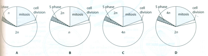
3 Adiploid cell undergoes a cell cycle including mitosis. Which diagram correctly shows the changes in chromosome number during interphase?
4 a Distinguish between the following terms:
i haploid and diploid
ii centromere and centriole
b Briefly explain what is meant by the following terms:
i autosome
ii homologous chromosomes
5 Thediagram shows three cells (labelled A, B and C) from a root tip which have been stained to show chromosomes.

b Describe what is happening at each stage.
6 Diagram 1 shows the life cycle of a simple plant known as a liverwort. Liverworts have two multicellular stages in life cycle: one is haploid and produces gametes; the other is diploid and produces spores.

a Copy diagram 1 and write 'mitosis' on one of the arrows in the life cycle where mitosis would take place.[1]
b Write 'meiosis' where meiosis would need to take place.[1]
c Explain why meiosis is needed in this life cycle.[3]
d Diagram 2 shows a cell of a liverwort plant dividing by mitosis. Only two of the many chromosomes are shown for simplicity.
i What stage of mitosis is shown? [1]
ii Is this cell haploid or diploid? Explain your answer.[3]
iii Draw prophase for the same cell (assume the cell has only two chromosomes, as in diagram 2).[1]
e Diagram 3 shows the same cell at telophase. The cell is beginning to divide and a new cell wall is forming, spreading out from the middle of the cell. Copy the diagram and add drawings of the chromosomes as they would appear at this stage.[1]
[Total: 7]
7 Microtubulesare tiny tubes made of protein subunits which join together. The protein is called tubulin. Colchicine isa natural chemical which binds to tubulin molecules, thus preventing the formation of microtubules.
a Why should the binding of colchicine to tubulin molecules interfere with the formation of microtubules? [2]
b What structure or structures involved in mitosis are made of microtubules? [2]
c When cells treated with colchicine are observed, the dividing cells are all seen to be in the same stage of mitosis. Suggestwith reasons the identity of this stage. [3]
[Total: 7]
Diagram1 shows chromosomes in the nucleus of a diploid cell.

a Draw the nucleus of a gamete produced from this cell. [1]
b What type of nuclear division would be used in the production of the gamete? [1]
c Draw a diagram to show what the nucleus would look like in anaphase of mitosis.[3]
Diagrams 2 and 3 below show the same diploid nucleus as in diagram1. However, the chromosomes have been shaded.

d State what the different types of shading represent in each nucleus.[2]
e Draw a karyogram based on the diploid nucleus shown in all 3 diagrams.[3]
[Total: 10]
9 Humans have 46 chromosomes in each body cell. Six cells are shown in the diagram below.
a Copy the diagram. For e ch cell, insert in the circle the number of chromosomes it contains. [3]
b What type of nuclear division takes place at X? [1]
[Total: 4]

1 D
2 B
3 D
4 a i haploid (cell or organism) has one set of chromosomes;
diploid has two sets of chromosomes;
ii centromere is region of a chromosome that holds two chromatids together;
centriole is an organelle;
found (in pairs) just outside nucleus;
microtubule organising centres/starting points for growing microtubules (for spindle);
b i a non-sex chromosome;
ii a pair of chromosomes that have the same structure;
same genes;
pair up during meiosis (forming a bivalent);
found in diploid cells;
5 a A anaphase; B prophase; C metaphase;
b Information for this answer can be found in Figure 5.10 on page 92 in the Coursebook.
Exam-style questions

b ‘meiosis’ label added to arrow between spore- producing stage and unicellular spores; [1]
c gamete-producing stage is haploid and spore- producing stage is diploid;
chromosome number would double every generation if no meiosis;
because life cycle includes sexual reproduction; haploid gametes fuse to form diploid zygote/when gametes fuse chromosome number, doubles/changes, from one set to two sets/gametes must be haploid and there is a diploid stage in the life cycle; [max. 3]
d i metaphase; [1]
ii haploid;
if it were diploid there would be, four pairs of chromatids/two long pairs of chromatids and two short pairs of chromatids, lined up on the equator;
chromosomes are lined up separately/not paired in homologous pairs as they would be in meiosis;3]
iii prophase drawing shows two single chromosomes, each with a centromere (not paired chromatids), ‘randomly’ distributed, surrounded by cell surface membrane but with no spindle; [1]
e a long and a short chromatid, each with a centromere, are shown inside each new nucleus; [1]
[Total: 11]
7 a microtubules are made out of tubulin molecules; the tubulin molecules stick together in a particular pattern to form the microtubules,
so the presence of colchicine would interfere with this; AW [2]
b spindle;
centrioles; [2]
c (held up in) prophase;
spindle cannot form (due to presence of colchicine);
so metaphase cannot occur;
metaphase, normally follows prophase/is next stage of mitosis; [max. 3]
[Total: 7]
8 a one long, one short and one hooked chromosome present inside a circle (nucleus); [1] b meiosis; [1]
c six chromatids about half way between equator and each pole (12 chromatids in all);
two long, two short, two hooked in each direction;
centromere leading for each chromatid; [3]
d in diagram 2, shading represent sets of chromosomes/one type of shading represents set of chromosomes from mother, other type of shading represents set of chromosomes from father; AW
in diagram 3, shading represent homologous pairs of chromosomes/differently numbered chromosomes; AW [2]
e only chromosomes drawn (no nuclear envelope); three separate homologous pairs drawn side by side;
pairs arranged in order of size, starting with largest; [3]
[Total: 10]
9 a body cells 46;sperm and egg 23;
zygote 46; [3]
b mitosis; [1]
[Total: 4]
A chromosome 10 micrometers long was found to contain 8.7 cm of DNA. What is the packing ratio of DNA in this chromosome? Show your working.
ReplyDeleteNo idea lol
ReplyDeleteI love your blog, useful info...
ReplyDelete2020 right here
Delete2020 right here
DeleteThis is the best blog ever
ReplyDeletei just want to say that you're handsome
ReplyDeleteThanks
ReplyDelete