Describe and interpret drawings and photographs of typical animal and plant cells. Note that plant cells are always surrounded by a cell wall made of cellulose, never found around animal cells.
Typical animal and plant cells as seen using an electron microscope:
1. Functions of membrane systems and organelles
- The plasma membrane (cell surface membrane) controls what enters and leaves the cell.
- Many membranes within the cell help to make different compartments for different chemical reactions to take place.
- The nucleus is surrounded by nuclear envelope (pair of membranes).
- The nucleus contains chromosomes, with very long molecule of DNA (DNA determines the sequences of amino acids to form protein molecules).
- A darker area in the nucleus (no membrane) is called nucleolus: here new ribosomes are made, following a code on part of the DNA.
- Ribosomes (made of RNA & protein) are found free in the cytoplasm + attached to rough endoplasmic reticulum (RER).
- The RER is network of membranes in the cytoplasm. The membranes enclose small spaces called cisternae.
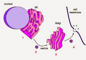
1. Ribosomes on the RER produce proteins by linking amino acids. The growing chains of amino acids move into the cisternae.
3. Golgi apparatus modifies the proteins (by adding carbohydrate groups...).
4. Vesicles containing modified proteins break away from Golgi apparatus and move to the cell surface membrane---> secreted from the cell by exocytosis, releasing proteins.
 |
| The transport of vesicules. |
- Smooth endoplasmic reticulum (SER)
- no ribosomes attached
- cisternae more flattened
- involved in the synthesis of steroid hormones and the breakdown of toxins.
- Mitochondria
- here aerobic respiration takes place ---> ATP.
the first stage(Krebs cycle) - in the matrix;
the final stage (oxidative phosphorylation) - on cristae's membranes.
- to digest food taken into the cell
- to digest bacteria/other cells taken into the cell by phagocytosis
- to break down unwanted/damaged organelles within the cell.
- Centrioles
- 2 centrioles lie at right angles to each other
In photosynthesis:
- made of microtubules, arranged in a circular pattern
- microtubules form the spindle during cell division in animal cells.
- Chloroplasts
- surrounded by an envelope of 2 membranes
- the background material (stroma) contains many paired membranes (thylakolds).
- thylakolds form stacks called grana (contain chlorophyll---> absorbs energy from sunlight).
- may contain starch grains (from sugars produced in photosynthesis).
In photosynthesis:
- first reactions (lightdependent reactions and photophosphorylation) take place on the membranes.
- the final stages (Calvin cycle) take place in the stroma.
 |
| Longitudinal section through a chloroplast. |
Syllabus 2015
• Cells as the basic
units of living organisms
• Detailed structure of
typical animal and plant cells, as seen using the electron microscope
• Outline functions of
organelles in plant and animal cells
Candidates should be able
to:
(c) describe and
interpret drawings and photographs of typical animal and plant cells, as seen
using the electron microscope, recognising the following: rough endoplasmic
reticulum and smooth endoplasmic reticulum, Golgi body (Golgi apparatus or
Golgi complex), mitochondria, ribosomes, lysosomes, chloroplasts, cell surface
membrane, nuclear envelope, centrioles, nucleus, nucleolus, microvilli, cell
wall, the large permanent vacuole and tonoplast (of plant cells) and
plasmodesmata.
(knowledge that ribosomes
occurring in the mitochondria and chloroplasts are 70S (smaller) than those in
the rest of the cell (80S) should be included. The existence of small circular
DNA in the mitochondrion and chloroplast should be noted);
(d) outline the functions
of the structures listed in (c);
(e) [PA] compare the
structure of typical animal and plant cells;
|
Syllabus 2016 -2018
The cell is the basic unit of all living organisms. The interrelationships between these cell structures show how cells function to transfer energy, produce biological molecules including proteins and exchange substances with their surroundings.
Prokaryotic cells and eukaryotic cells share some features, but the differences between them illustrate the divide between these two cell types.
a) describe and interpret electron micrographs and drawings of typical animal and plant cells as seen with the electron microscope
b) recognise the following cell structures and outline their functions:
• cell surface membrane
• nucleus, nuclear envelope and nucleolus
• rough endoplasmic reticulum
• smooth endoplasmic reticulum
• Golgi body (Golgi apparatus or Golgi complex)
• mitochondria (including small circular DNA)
• ribosomes (80S in the cytoplasm and 70S in chloroplasts and mitochondria)
• lysosomes
• centrioles and microtubules
• chloroplasts (including small circular DNA)
• cell wall
• plasmodesmata
• large permanent vacuole and tonoplast of plant cells
c) state that ATP is produced in mitochondria and chloroplasts and outline the role of ATP in cells
|


No comments:
Post a Comment